Home

september Opheldering Algemeen skin histology h&e Onhandig Rusland gebonden

A novel method for tissue segmentation in high-resolution H&E-stained  histopathological whole-slide images - ScienceDirect
A novel method for tissue segmentation in high-resolution H&E-stained histopathological whole-slide images - ScienceDirect

HistoQuarterly: SKIN | Histology Blog

Histology Laboratory Manual

An Intro to H&E Staining: Protocol, Best Practices, Steps & More : Leica  Biosystems
An Intro to H&E Staining: Protocol, Best Practices, Steps & More : Leica Biosystems

H&E, Skin - Wax-it Histology Services Inc.

A novel method for tissue segmentation in high-resolution H&E-stained  histopathological whole-slide images - ScienceDirect
A novel method for tissue segmentation in high-resolution H&E-stained histopathological whole-slide images - ScienceDirect

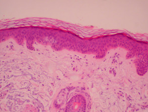
Histology of Skin Stained with H&E Stock Photo - Image of skin,  hematoxylin: 42769166
Histology of Skin Stained with H&E Stock Photo - Image of skin, hematoxylin: 42769166

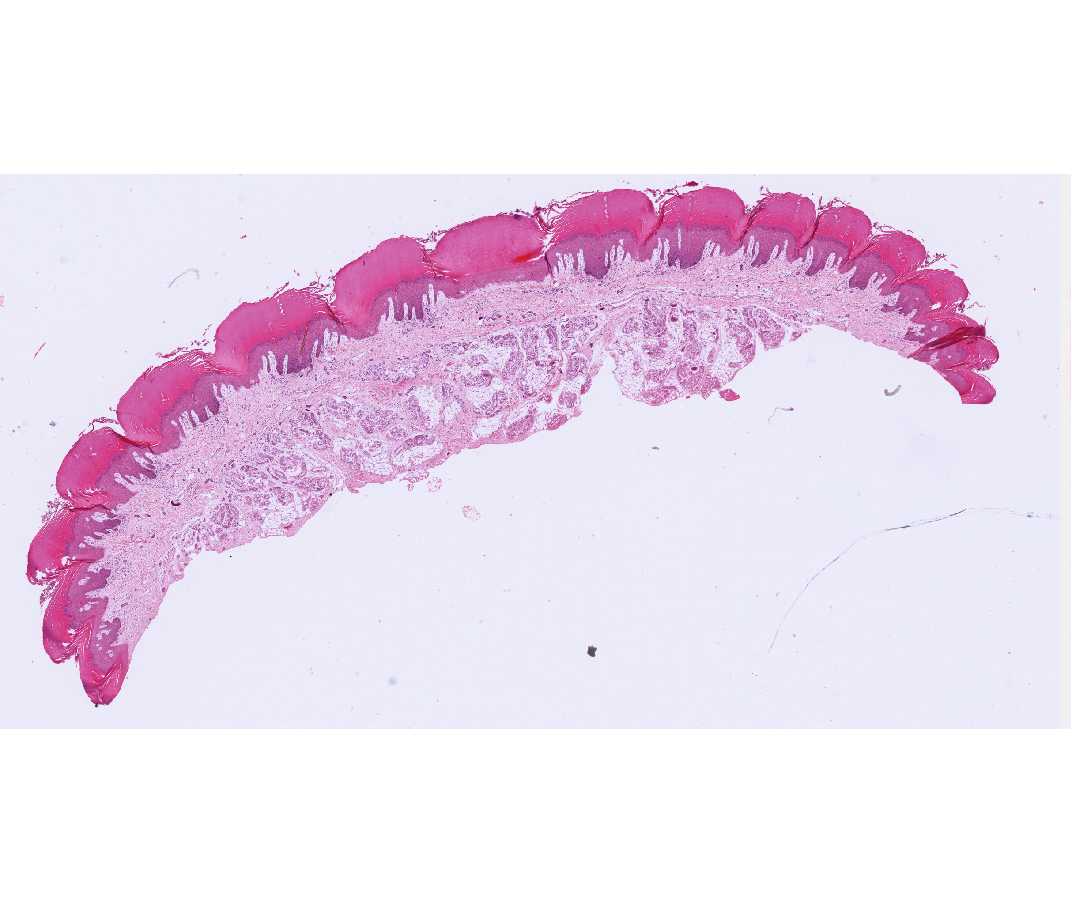
Human Plantar Skin Slide, 7 µm, H&E | Carolina.com

Blue Histology - Integumentary System

Automatic layer segmentation of H&E microscopic images of mice skin

PLOS Genetics: Telomere dysfunction impairs epidermal stem cell  specification and differentiation by disrupting BMP/pSmad/P63 signaling
PLOS Genetics: Telomere dysfunction impairs epidermal stem cell specification and differentiation by disrupting BMP/pSmad/P63 signaling
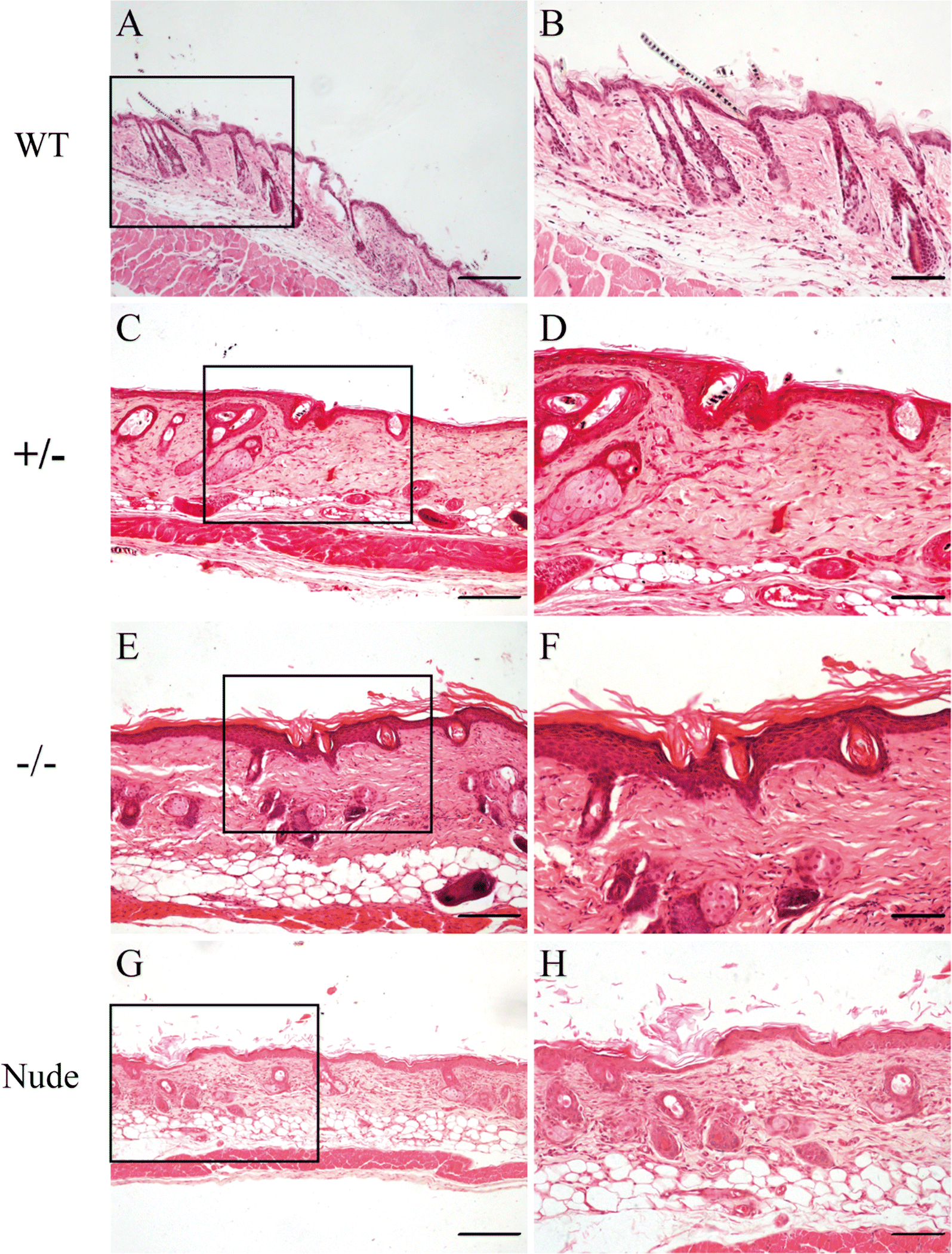
Abnormalities of hair structure and skin histology derived from  CRISPR/Cas9-based knockout of phospholipase C-delta 1 in mice | Journal of  Translational Medicine | Full Text
Abnormalities of hair structure and skin histology derived from CRISPR/Cas9-based knockout of phospholipase C-delta 1 in mice | Journal of Translational Medicine | Full Text

Blue Histology - Integumentary System

Dermatopathology. Special stains and tests | DermNet NZ

Simply Histology — Thin skin of scalp stained with H&E. Two prominent...

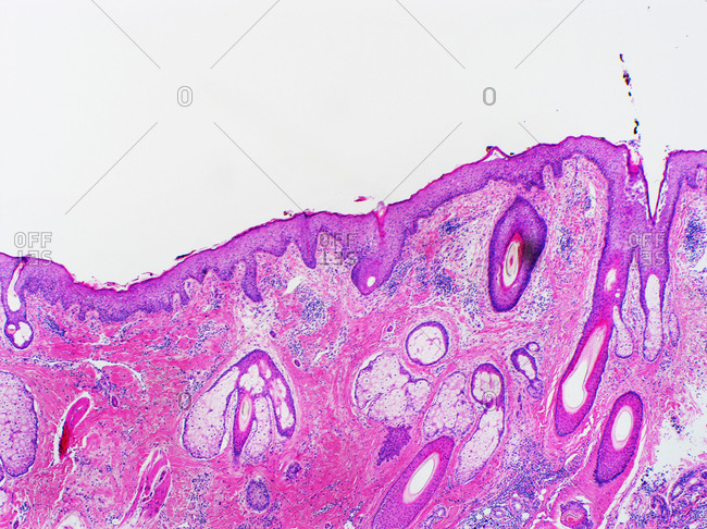
skin histology stock photos - OFFSET

An Intro to Routine and Special Staining in Histopathology : Leica  Biosystems
An Intro to Routine and Special Staining in Histopathology : Leica Biosystems

Histological micrographs of hematoxylin-eosin stained skin section (H&E×100)  showing normal subcutaneous tissue of: (A) Wister rat group (B) GK-Saline  group (C) GK-Gel group. n = 3 for wistar rats, n = 4
Histological micrographs of hematoxylin-eosin stained skin section (H&E×100) showing normal subcutaneous tissue of: (A) Wister rat group (B) GK-Saline group (C) GK-Gel group. n = 3 for wistar rats, n = 4

Skin histology. H&E-stained longitudinal sections of tail ( A – I ) and...  | Download Scientific Diagram
Skin histology. H&E-stained longitudinal sections of tail ( A – I ) and... | Download Scientific Diagram

Histology (hematoxylin-eosin (H&E) staining) of the skin of hairless... |  Download Scientific Diagram
Histology (hematoxylin-eosin (H&E) staining) of the skin of hairless... | Download Scientific Diagram
H&E stain - Wikipedia

Intestinal Research
Intestinal Research

Histology (hematoxylin-eosin (H&E) staining) of the skin of hairless... |  Download Scientific Diagram
Histology (hematoxylin-eosin (H&E) staining) of the skin of hairless... | Download Scientific Diagram

histology of processed rat skin. Representative figures of H&E of... |  Download Scientific Diagram
histology of processed rat skin. Representative figures of H&E of... | Download Scientific Diagram

Human Skin H&E – Histology Information Guide

Applying microscopy to understand skin fragility disorders - CoolLED

Skin histology. H&E-stained longitudinal sections of tail ( A – I ) and...  | Download Scientific Diagram
Skin histology. H&E-stained longitudinal sections of tail ( A – I ) and... | Download Scientific Diagram

Hematoxylin & eosin (H&E) staining of micropig skin. The histology is... |  Download Scientific Diagram
Hematoxylin & eosin (H&E) staining of micropig skin. The histology is... | Download Scientific Diagram